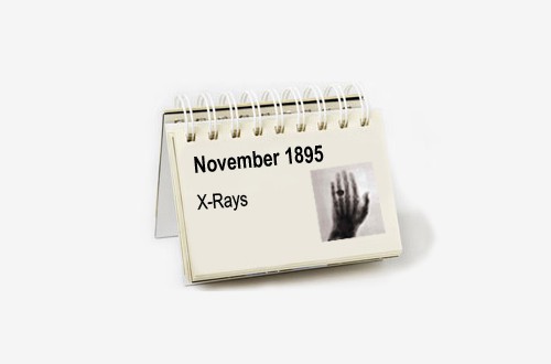
“I have seen my death”. These were the words of Anna Bertha Röntgen upon being the first person in the world to see her own skeleton while it remained safely encased in muscle and skin.
For Anna Bertha Röntgen was the wife of German scientist Wilhelm Conrad Röntgen and in 1895 she became the subject of his newly discovered and still inexplicable X-ray radiation that made visible the invisible.
Her words were suitably dramatic, capturing a dark 19th century fascination with fledgling science, anatomy, death and rebirth. But, in perhaps more practical terms, rather than seeing death, what Anna actually saw was the bone structure of her left hand and the wedding ring on her finger.
Wilhelm would have seen these too. And he would also have seen something beyond the tangible, although not the morbid evidence of human mortality that his wife observed.
Instead, Wilhelm saw possibilities beyond what science had previously thought possible and something that would prove to be one of the greatest advances in medical history.
As with many other healthcare breakthroughs, the discovery of the X-ray has been put down to some form of happy accident.
The scientific environment at the time provides leverage to a story of all the right parts falling into place at the right time, rather than one of some freak deus ex machina turning the world on its head.
For, as described by Dr Howard Markel, director of the Center for the History of Medicine at the University of Michigan, “the phenomenon of electricity and electrons was all the rage”.
Scientists across the world were playing with this still infant technology, and one of the most imaginative was Wilhelm Röntgen.
The traditional story describes how Röntgen was experimenting with a Crookes tube – an early device that was able to create streams of electrons , known as cathode rays, in a vacuum when a high voltage was applied between its two electrodes.
Up to that point, the tubes had been a curiosity for inventive minds rather than holding a specific function, but on November 8, 1895, Röntgen made a very practical discovery using a tube that featured a thin window that allowed some of the rays to escape.
Two stories currently circulate as to exactly what happened on this day, but either scenario takes into account agreeable circumstances.
One account claims that Röntgen left a Crookes tube on a book while out for lunch. Fortuitously, the book happened to be resting on a piece of photographic film, while in the book there rested a metal key.
Upon returning to his laboratory, Röntgen discovered the image of the key on the film – the world’s first X-ray photograph.
Story two also depends on an aspect of chance, with Röntgen alone at night at the German University of Würzburg conducting an experiment to see what happened when the currents created by the Crookes tube passed through certain gases.
As he carried out this task, he noticed that a nearby screen began to glow, inspiring him to test the rays on other materials. This led to the discovery that the beams could pass through certain materials but not others, and were able to leave an imprint of these resistant objects on photographic film.
In any case, early subjects included weights and pieces of metal, before Röntgen convinced his wife to place her hand in the path of these mysterious rays just before Christmas in 1895.
Still uncertain of just what he had unearthed, Röntgen names his discovery the ‘X-ray’, publishing his initial findings at the end of 1895.
With such an astonishing breakthrough that easily captured the public’s imagination, it was mere weeks before news spread around the world, reaching scientists, physicians and anyone else interested in seeing ‘death’.
The potential in healthcare was obvious, and uptake was quick, with the first recorded use of an X-ray for clinical use in January 1896 when English physicist John Hall-Edwards took an image of a needle stuck in someone’s hand.
Just one month later on February 14, 1896, Hall-Edwards became the first person to use an X-ray as a diagnostic tool before an operation.
Hall-Edwards unfortunately became victim to the miraculous device when he developed X-ray dermatitis due to exposure to the radiation and had to have his left arm amputated.
Other scientists, such as Thomas Edison’s assistant Clarence Dally, were also harmed, with Dally dying of skin cancer in 1904.
The lack of awareness of the danger of X-rays continued through the 20th century as shoe stores used devices to let customers see bones in their feet to understand the effect of tight shoes.
Eventually, safety caught up with the science, and X-rays became standard healthcare practice throughout the world, influencing the next generation of diagnostic devices, such as CAT scans, MRIs and ultrasound.
For his contribution, Röntgen was presented with the Nobel Prize in physics in 1901, and perhaps more excitingly for a scientist, the element roentgenium was named in his honour in 2004 – more than a century after his greatest discovery.


